
Raman Lab
Raman Lab
Leader: Mr Paul Schreiner
Deputy: Ms Lu Chen
Our Raman Laboratory for the characterisation of materials. Consulting on Raman spectroscopy applications is also offered.
Raman Spectroscopy is the inelastic scattering of photons by phonons. It is a simple, fast, non-destructive characterisation tool widely used in basic and applied research in natural sciences and industries. Temperature controlled stages (Oxford Instrument and Linkam) are available for temperature dependent measurements in the range of -196 and 600 ℃. For training on the equipment and access to the lab, or for any general training, please contact the Lab Leader or their deputy.
While there is a range of devices in the Raman, several of them require booking before use.
The booking system can be found here.
AttoCube He-free CryogenicPrincipal User: Dr Yang Li Automated top-loading cryostat with variable temperature & superconducting magnet - closed-cycle & low vibration cryostat platform - no liquid helium required & enables SPM - automated temperature and magnetic field control - fast parameter change via touchscreen - variable temperature (1.65 .. 300 K) @ full field - transport measurements & LT-SPM | 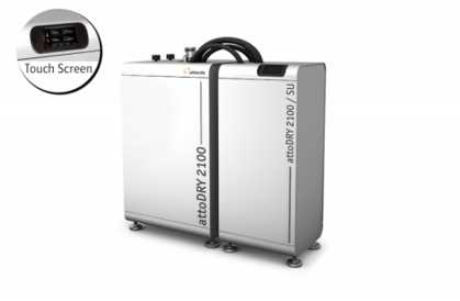 |
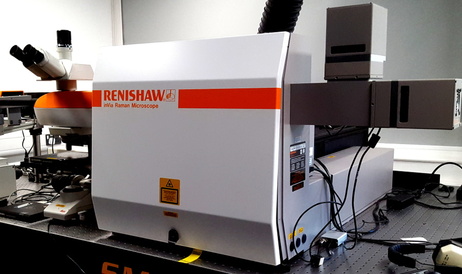
Renishaw RM1000Principal User: Mr Paul Schreiner | 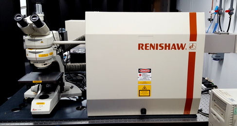 |
Renishaw InViaPrincipal User: Mr Paul Schreiner The highly efficient optical design gives you the best Raman data, from minute traces of material and large volumes. Apply this power to your experiments, running measurements such as:
|  |
Horiba LabRam HR800Principal User: Mr Paul Schreiner HORIBA Jobin Yvon's LabRAM 800HR for example is a unique bench-top system that combines micro Raman and photoluminescence (PL) spectroscopy in a single instrument. This means that academic researchers and manufacturers of silicon and group III and V semiconducting materials can measure both the vibrational characteristics of the molecules and their electronic structure quickly and conveniently. This yields a much greater depth of information than is possible from just a single analysis. The instrument incorporates an integral confocal Raman / PL microscope, making it possible to detect discontinuities in crystal growth, homogeneity, composition, contamination, dopant effects and regions of stress on the micron scale. The user can switch easily between Raman and PL spectroscopy at the same point on the sample without any need for optical adjustment | 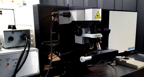 |
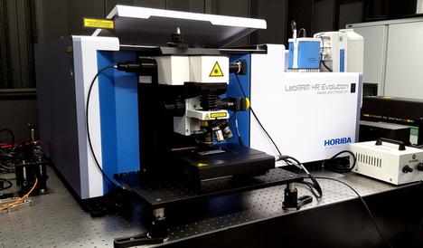
Horiba LabRAM EvolutionPrincipal User: Mr Paul Schreiner The LabRAM HR Evolution Raman microscopes are ideally suited for both micro and macro measurements, and offer advanced confocal imaging capabilities in 2D and 3D. The true confocal Raman microscope enables the most detailed images and analyses to be obtained with speed and confidence. With guaranteed high performance and intuitive simplicity, the LabRAM HR Evolution is the ultimate instrument for Raman spectroscopy. They are widely used for standard Raman analysis, photoluminescence (PL), tip enhanced Raman scattering (TERS) and other hybrid methods.
|  |
M2 laser setupPrincipal User: Mr Paul Schreiner Deputy User: Ms Lu Chen descriptionxxxxxxx |
When in the Raman laboratory, please follow the local rules, downloadable here.